

 Verified patient reviews on Doctify & Top Doctors →

Professional memberships
Providing care in London centres
- LondonHarley Street · Canary Wharf · Battersea
- EssexChelmsford
His verified patient reviews on Top Doctors, collected and checked by Top Doctors itself.
Read his Top Doctors reviews ↗Ready to talk it through?
Call the practice office, email us at Masannat.Admin@LIPS.org.uk, or send a request and the team will call you back.
One clinic, every stage of breast care
From benign lumps to complex reconstruction — choose an area to read more about it.
Breast lumps & benign conditions
A one-stop clinic assessing breast pain, lumps, cysts, nipple changes and infections — most findings turn out to be benign, and same-visit reassurance is the goal.
- Breast cysts & fibroadenomas
- Breast pain & nipple discharge
- Breast infection & abscess
- Benign lump excision, duct excision
- Nipple eczema, armpit mass, axillary lymph nodes
Surgeon, academic, and patient advocate

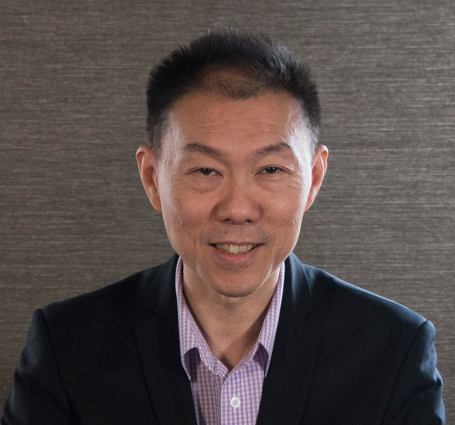
Dr Yazan Masannat is a Consultant Oncoplastic and Reconstructive Breast Surgeon in London, practising on Harley Street, in Canary Wharf and in Battersea, and Visiting Associate Professor at Anglia Ruskin University. He specialises in the diagnosis and treatment of breast cancer, oncoplastic breast-conserving surgery, breast reconstruction and benign breast conditions, with a one-stop clinic for new symptoms.
He has published more than 100 peer-reviewed papers, holds a National Clinical Impact Award, and is the founder and editor of iBreastBook, a free international education platform for breast surgeons. He is approved by all major insurers including Bupa, AXA Health, Aviva, Cigna, WPA and Vitality.
Appointments & roles
Meet the MDT team
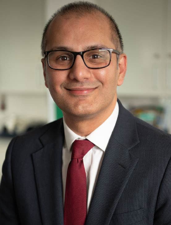
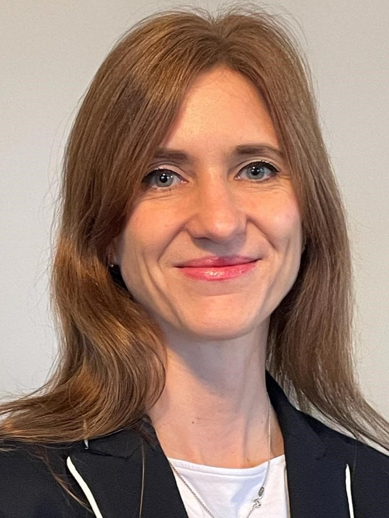
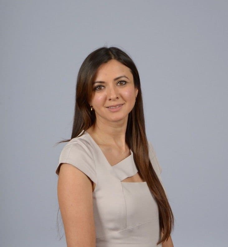
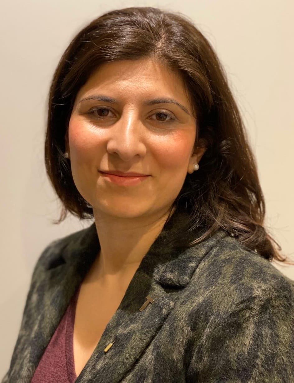
Breast care works best as a team. Dr Masannat works closely with this multidisciplinary group of oncologists, plastic surgeons and radiologists across London.







Creator of iBreastBook
iBreastBook is a free, global education platform for breast surgery, founded and edited by Dr Yazan Masannat with Wafa Ghattas. It brings together expert-led webinars, teaching resources and an international community of breast surgeons, radiologists, oncologists and trainees. Its webinar series, started during the COVID pandemic, is now an internationally recognised teaching programme.
100+ peer-reviewed publications
A selection of Dr Masannat's most recent research. The full list is on his research profiles:



The Prognostic Significance of the Serum Inflammatory Marker Ratios, Neutrophil-Lymphocyte, Platelet-Lymphocyte and Monocyte-Lymphocyte in Male Breast Cancer.
Cancer Invest. 2025 Jul;43(6):391-398.Read paper ↗2025A core outcome set for locoregional treatment reporting in neoadjuvant systemic breast cancer treatment trials.
NPJ Breast Cancer. 2025 Oct 28;11(1):116.Read paper ↗2025Long term outcomes of breast primary sarcomas and malignant phyllodes tumors: 20 years observational analysis of the BEAM study group.
Eur J Surg Oncol. 2025.Read paper ↗2025Consensus process to agree upon surgical quality assurance processes within a pragmatic, multicentre randomised clinical trial comparing targeted axillary dissection and axillary node clearance: TADPOLE-TOGETHER.
BMJ Open. 2025.Read paper ↗2024Residual microcalcifications after neoadjuvant systemic therapy for early breast cancer: implications for surgical planning and long-term outcomes.
Eur J Surg Oncol. 2024 Oct 22.Read paper ↗2024Field cycling imaging to characterise breast cancer at low and ultra-low magnetic fields below 0.2 T.
Commun Med (Lond). 2024 Oct 30;4(1):221.Read paper ↗2024Revisiting surgical margins for invasive breast cancer patients treated with breast conservation therapy — evidence for adopting a 1mm negative width.
Eur J Surg Oncol. 2024 Oct;50(10):108573.Read paper ↗2024Oncoplastic breast surgery in Europe.
Gland Surg. 2024 Feb 29;13(2):248-256.Read paper ↗2023The Role of Social Media in Breast Cancer Care and Survivorship: A Narrative Review.
Breast Care (Basel). 2023 Jun;18(3):203-208.Read paper ↗2023Chest wall perforator flaps for breast reconstruction: international survey on attitudes and training needs.
Br J Surg. 2023 Jul 17;110(8):966-972.Read paper ↗2020Therapeutic mammaplasty is a safe and effective alternative to mastectomy with or without immediate breast reconstruction.
Br J Surg. 2020 Jun;107(7):832-844.Read paper ↗2013Challenges in the management of pleomorphic lobular carcinoma in situ of the breast.
Breast. 2013 Apr;22(2):194-196.Read paper ↗Videos from Dr Masannat
Short explainers with Dr Masannat on breast health, diagnosis and treatment.
Trusted information, in one place
Information from established breast care and cancer support charities about symptoms, tests, treatment and recovery. It can help you prepare questions for your appointment.
137 resources
Know your breasts quick guide
A brief introduction to checking your breasts, recognising changes and seeking advice.
View booklet ↗Breast Cancer Now · BookletYour breast clinic appointment
Explains what happens at a breast clinic appointment and the tests that may be offered.
View booklet ↗Breast Cancer Now · Online guideMammogram and breast ultrasound
Explains what mammography and breast ultrasound involve during assessment.
Read guide ↗Breast Cancer Now · Online guideBreast biopsy
Explains breast biopsy, how samples are taken and what patients can expect.
Read guide ↗Breast Cancer Now · Online guideBreast lumps and benign (not cancer) breast conditions
An overview of non-cancerous breast conditions and links to more specific information.
Read guide ↗Macmillan Cancer Support · BookletUnderstanding breast cancer
A broad introduction to breast cancer, diagnosis, treatment and emotional and practical concerns.
View booklet ↗Breast Cancer Now · BookletYour operation and recovery
Explains preparation for breast cancer surgery, the hospital stay and recovery at home.
View booklet ↗Breast Cancer Now · BookletBreast reconstruction
An overview of reconstruction choices, benefits, limitations and recovery to support an informed decision.
View booklet ↗Breast Cancer Now · BookletExercises after breast cancer surgery
Illustrated exercises to help restore shoulder and arm movement at different stages after surgery.
View booklet ↗Breast Cancer Now · BookletReducing the risk of lymphoedema
Explains lymphoedema and steps that may help reduce its risk after breast cancer treatment.
View booklet ↗Breast Cancer Now · BookletAfter breast cancer treatment: what now?
Explains follow-up after hospital treatment, changes to report and adjusting emotionally when treatment ends.
View booklet ↗Macmillan Cancer Support · BookletHelp with the cost of cancer
Explains financial support for people affected by cancer and how to find help with claiming it.
View booklet ↗Common questions, answered plainly
These answers provide general information and cannot replace an individual consultation. Breast symptoms are common and most are not caused by cancer, but any new or unusual change should be assessed. If you are seriously unwell or have a medical emergency, call 999 or attend your nearest emergency department.
No. Most breast lumps are benign (not cancer), with common causes including cysts, fibroadenomas and normal hormonal breast changes. However, it is not possible to confirm the cause by touch alone, so any new lump or persistent thickening should be assessed clinically and may need imaging.
Read more: Breast lumps & benign conditions →Arrange an assessment for any new or unusual change, including a lump or swelling in the breast, upper chest or armpit; skin dimpling or redness; a change in breast size or shape; a newly inverted nipple; a nipple rash or crusting; or spontaneous nipple discharge. Do not wait for the change to become painful or for another symptom to develop.
Breast pain on its own is rarely caused by cancer. It is often hormonal or related to the chest wall, muscles, cysts, medication or an ill-fitting bra. New, persistent or localised pain—particularly when accompanied by a lump, skin change, nipple change or swelling—should still be assessed.
A one-stop clinic brings together consultation, clinical examination and breast imaging during the same visit wherever possible. If an abnormality needs tissue sampling, a needle biopsy can often be performed at the same appointment. Imaging findings may be available on the day, but biopsy results take longer, usually within a few days and up to a week in most cases.
Triple assessment combines three elements: clinical assessment, imaging and a needle biopsy when indicated. The results are interpreted together because no single test answers every question. Not every patient needs all three elements—for example, a biopsy is only performed when there is an area that requires sampling.
In Dr Masannat’s one-stop clinics, everyone aged 40 or over who attends with a breast concern will have a mammogram as part of the assessment. Ultrasound is frequently added because it provides complementary information, particularly when assessing a specific lump or focal symptom. In patients under 40, ultrasound is usually the first imaging test, with mammography added if clinically indicated.
A core biopsy uses a needle to take small samples of tissue, usually under ultrasound or mammographic guidance and with local anaesthetic. You may feel pressure or brief discomfort, and bruising or tenderness can last for a few days. The tissue is examined by a specialist breast pathologist to establish the diagnosis.
For appropriately selected early breast cancers, breast-conserving surgery followed by radiotherapy provides at least equivalent long-term cancer control and survival to mastectomy. The best option depends on the extent of disease, breast-to-tumour size ratio, genetic risk, previous treatment, likely radiotherapy and your preferences.
Read more: Breast cancer →Oncoplastic surgery combines cancer removal with plastic-surgical reshaping techniques. It can allow a larger area to be removed while preserving or improving breast shape and may include a breast reduction or lift. The opposite breast may sometimes be adjusted for balance, either at the same operation or later.
Read more: Oncoplastic surgery →The breast shape can be rebuilt using an implant, your own tissue (a flap), or a combination of the two. Common tissue-based options use skin and fat from the abdomen (DIEP, SIEA), buttocks (SGAP, IGAP), thigh (TUG, PAP) or another donor area. Each option has different operating times, recovery, scars, risks and long-term behaviour.
Read more: Breast reconstruction →Recovery varies considerably with the operation. Many people resume light daily activities within days after smaller breast operations, while mastectomy and reconstruction require longer. Wounds often heal over several weeks, but swelling, altered sensation and tiredness can take several months to settle.
You can submit a booking request through the website or contact the practice team by telephone or email. The team will confirm the most suitable clinic, appointment time and whether imaging should be arranged for the same visit.
Read more: Request an appointment →No. Most breast lumps are benign (not cancer), with common causes including cysts, fibroadenomas and normal hormonal breast changes. However, it is not possible to confirm the cause by touch alone, so any new lump or persistent thickening should be assessed clinically and may need imaging.
Read more: Breast lumps & benign conditions →Arrange an assessment for any new or unusual change, including a lump or swelling in the breast, upper chest or armpit; skin dimpling or redness; a change in breast size or shape; a newly inverted nipple; a nipple rash or crusting; or spontaneous nipple discharge. Do not wait for the change to become painful or for another symptom to develop.
Breast pain on its own is rarely caused by cancer. It is often hormonal or related to the chest wall, muscles, cysts, medication or an ill-fitting bra. New, persistent or localised pain—particularly when accompanied by a lump, skin change, nipple change or swelling—should still be assessed.
Most nipple discharge is benign, but new or spontaneous discharge should be assessed, particularly if it comes from one breast or one duct, is blood-stained or clear, or is associated with a lump or nipple change. Milky discharge from both breasts can be hormonal or medication-related and may require assessment by your GP as well as a breast specialist.
Some people have naturally inverted nipples. A nipple that has recently pulled in, changed direction or become distorted—especially on one side—needs assessment. Infection, duct changes and cancer are among the possible causes, and examination and imaging help distinguish between them.
An armpit lump may be an enlarged lymph node, a skin cyst, inflammation, accessory breast tissue or another benign condition. Occasionally it can relate to breast cancer or another illness. A persistent, enlarging or unexplained armpit lump should be examined.
Yes, although it is uncommon. Men should seek assessment for a new chest or breast lump, nipple discharge, nipple inversion, skin ulceration or swelling in the armpit. The same principle applies: most symptoms will not be cancer, but they should be checked.
Yes. Many breast changes during pregnancy and breastfeeding are benign, but a persistent lump, redness that does not improve, bloody discharge or another new change should not be ignored. Ultrasound is commonly the first imaging test, and mammography or biopsy can be performed safely when clinically necessary.
These symptoms may be caused by mastitis or an abscess and should be assessed promptly, particularly if you feel feverish or unwell. Treatment may include antibiotics and, if an abscess is present, drainage that is usually done under ultrasound scan guidance. Redness or swelling that does not settle with appropriate treatment needs further investigation, as a type of breast cancer called inflammatory breast cancer usually presents with a red, swollen breast.
A one-stop clinic brings together consultation, clinical examination and breast imaging during the same visit wherever possible. If an abnormality needs tissue sampling, a needle biopsy can often be performed at the same appointment. Imaging findings may be available on the day, but biopsy results take longer, usually within a few days and up to a week in most cases.
Triple assessment combines three elements: clinical assessment, imaging and a needle biopsy when indicated. The results are interpreted together because no single test answers every question. Not every patient needs all three elements—for example, a biopsy is only performed when there is an area that requires sampling.
The consultation usually begins with a discussion of your symptoms, previous breast problems, medical and family history, medication and any previous breast surgery. Dr Masannat will then examine both breasts and, where appropriate, the lymph nodes. He will explain whether imaging or another test is recommended and discuss the findings and next steps with you.
You are welcome to bring a partner, relative or friend. This can be particularly helpful if you are anxious, attending for biopsy results or discussing treatment options. A chaperone can also be provided for the examination.
In Dr Masannat’s one-stop clinics, everyone aged 40 or over who attends with a breast concern will have a mammogram as part of the assessment. Ultrasound is frequently added because it provides complementary information, particularly when assessing a specific lump or focal symptom. In patients under 40, ultrasound is usually the first imaging test, with mammography added if clinically indicated.
MRI is a specialist test and dense breast tissue alone does not automatically mean that an MRI is needed. However, in certain clinical presentations, MRI can be discussed as a further imaging tool when additional information is required. It may also be recommended for selected people at high inherited risk, to clarify the extent of a known cancer, to assess difficult or conflicting findings, or to evaluate breast implants. The decision is made in the context of the clinical examination, mammography, ultrasound and the individual clinical question.
Contrast-enhanced mammography combines mammography with an iodine-based contrast injection to show areas with increased blood supply. It can provide additional information in selected cases, including some situations in which MRI would otherwise be considered. It is not needed for everyone and may not be suitable if you have a significant contrast allergy or certain kidney problems.
Mammography is an important and effective test, but no imaging test detects every breast cancer. Accuracy can be affected by breast density and by the type and position of an abnormality. For a new symptom, mammography should not be considered in isolation: clinical examination, ultrasound and biopsy may also be required.
A core biopsy uses a needle to take small samples of tissue, usually under ultrasound or mammographic guidance and with local anaesthetic. You may feel pressure or brief discomfort, and bruising or tenderness can last for a few days. The tissue is examined by a specialist breast pathologist to establish the diagnosis.
No. Biopsies are performed to identify the nature of an abnormality, and many biopsy results are benign. The purpose is to replace uncertainty with a tissue diagnosis and ensure that any treatment or follow-up recommendation is appropriate.
A very small metal marker may be left at the biopsy site so the area can be found again on future imaging or at surgery. It is usually not felt, does not set off airport security systems and is designed to remain safely in the breast. Most modern breast marker clips are MRI compatible, although you should always tell the radiology team about any implant or device before a scan.
Mammogram and ultrasound findings are often discussed during the same visit. Biopsy results usually take several working days and can take longer if additional laboratory tests are needed. The team will tell you how and when your results will be communicated rather than leaving you to arrange this yourself.
The clinical, imaging and biopsy findings must be consistent, and all biopsies are discussed in a multidisciplinary team meeting to make sure the findings agree. If they are not, the case may need further imaging, repeat biopsy and sometimes surgical excision. A reassuring scan should not simply override a persistent suspicious clinical finding.
Ductal carcinoma in situ (DCIS) is a non-invasive breast cancer in which abnormal cells are contained within the milk ducts and have not invaded the surrounding breast tissue. It is usually treated with surgery, sometimes followed by radiotherapy, to prevent recurrence or progression to invasive cancer. The recommended treatment depends on its size, grade and distribution.
Read more: Breast cancer →Grade describes how abnormal the cancer cells look under the microscope and how quickly they are likely to grow. Stage describes the size and extent of the invasive cancer, including whether lymph nodes or other parts of the body are involved. Both help the multidisciplinary team recommend treatment, but they answer different questions.
These are biomarkers tested on invasive breast cancers. Oestrogen receptor (ER) and progesterone receptor (PR) results help determine whether the tumour is hormone sensitive and if endocrine treatment is likely to help, while HER2 results determine whether HER2-targeted medicines may be beneficial; these are usually given with chemotherapy. They are only part of the overall treatment decision.
Invasive lobular cancer begins in the milk-producing lobules and can grow in a more diffuse pattern than the commoner invasive ductal/no-special-type cancer. It may be less conspicuous on examination or mammography, so additional imaging is sometimes considered. Treatment is individualised in the same multidisciplinary way as for other invasive cancers.
The MDT brings together breast surgeons, radiologists, pathologists, medical and clinical oncologists and specialist breast care nurses. They review the imaging, pathology and relevant clinical information and agree evidence-based recommendations. The final treatment decision is made with you after your options, priorities and preferences have been discussed.
Some patients benefit from systemic treatment before surgery—known as neoadjuvant treatment—to shrink the cancer, treat the whole body early and assess how the tumour responds. This is more commonly considered for some HER2-positive, triple-negative or larger cancers, but it depends on the tumour biology and your individual circumstances.
Genetic assessment may be recommended depending on your age at diagnosis, cancer type, ancestry and family history of breast, ovarian, pancreatic, prostate or certain other cancers. Testing can influence your own treatment and screening and may have implications for relatives. It should be accompanied by appropriate counselling about what the result may—and may not—mean.
Yes. A second opinion can be helpful when the diagnosis is complex, you have been offered several surgical options, reconstruction is being considered or you want greater confidence before treatment. Bring or arrange access to your imaging, pathology reports, clinic letters and proposed treatment plan so the review is meaningful.
The main breast operations are breast-conserving surgery, which removes the cancer with a rim of surrounding tissue, and mastectomy, which removes the breast. Oncoplastic techniques may be used to reshape the breast during breast-conserving surgery, and reconstruction may be offered with or after mastectomy. Surgery to assess or treat the lymph nodes may also be required.
Read more: Breast cancer →For appropriately selected early breast cancers, breast-conserving surgery followed by radiotherapy provides at least equivalent long-term cancer control and survival to mastectomy. The best option depends on the extent of disease, breast-to-tumour size ratio, genetic risk, previous treatment, likely radiotherapy and your preferences.
Read more: Breast cancer →This operation removes the cancer with a margin of surrounding breast tissue while retaining the rest of the breast. Radiotherapy is usually recommended afterwards, although there are selected exceptions. The tissue is examined to confirm the diagnosis, tumour features and whether the margins are clear.
If cancer cells are too close to or reach the edge of the removed tissue, further surgery may be recommended. This may be a re-excision to remove more tissue or, less commonly, a mastectomy. The decision depends on the type and extent of disease, the expected cosmetic result and your preferences.
Oncoplastic surgery combines cancer removal with plastic-surgical reshaping techniques. It can allow a larger area to be removed while preserving or improving breast shape and may include a breast reduction or lift. The opposite breast may sometimes be adjusted for balance, either at the same operation or later.
Read more: Oncoplastic surgery →A mastectomy aims to remove as much breast tissue as possible, but no operation can remove every breast cell; in practice, approximately 90–95% of the breast tissue is removed, never 100%. It may be advised when disease is extensive relative to breast size, affects several widely separated areas, clear margins cannot be achieved, radiotherapy is unsuitable, cancer has recurred after previous breast-conserving treatment, or risk-reducing surgery is chosen because of a high inherited risk. Personal preference is important, but a more extensive operation does not provide better survival.
Nipple-sparing mastectomy is possible for selected patients when it is oncologically safe and the blood supply to the skin and nipple is likely to be adequate. Suitability depends on the tumour position and extent, breast shape, previous surgery or radiotherapy and factors that affect wound healing. Even when preserved, nipple sensation commonly changes or is lost.
An impalpable cancer can be marked before surgery using a wire, magnetic seed, radar reflector or another image-guided localisation device. This enables the surgeon to target the correct area while limiting removal of unnecessary tissue. The exact method depends on the hospital and the imaging findings.
A sentinel node biopsy identifies and removes the first lymph node or nodes most likely to receive drainage from the breast. A tracer is used to locate them, and they are examined for cancer cells. This provides staging information with a lower risk of arm complications than removing many lymph nodes.
Not always. Depending on the amount of cancer in the node, the breast operation, planned radiotherapy, tumour biology and any treatment already given, further axillary surgery may be unnecessary. The MDT may recommend radiotherapy, axillary clearance or no additional axillary treatment.
Axillary clearance removes a larger group of lymph nodes from the armpit. It is used selectively when the expected benefit outweighs risks such as numbness, shoulder stiffness and lymphoedema. Your team will explain why it is recommended and how these risks can be reduced and managed.
No. Reconstruction is a personal choice, not a requirement. Some patients choose immediate or delayed reconstruction, while others prefer a flat closure or an external breast prosthesis. You should receive balanced information and time to consider the options that are medically suitable for you.
Immediate reconstruction is performed during the same operation as the mastectomy, keeping the breast skin and sometimes the nipple. Delayed reconstruction is carried out months or years later after a flat mastectomy. Timing depends on the cancer treatment plan, possible radiotherapy, your health, the reconstructive options available and your preference.
The breast shape can be rebuilt using an implant, your own tissue (a flap), or a combination of the two. Common tissue-based options use skin and fat from the abdomen (DIEP, SIEA), buttocks (SGAP, IGAP), thigh (TUG, PAP) or another donor area. Each option has different operating times, recovery, scars, risks and long-term behaviour.
Read more: Breast reconstruction →Implant reconstruction usually involves a shorter operation and no distant donor-site scar, but implants are not lifetime devices and may require future surgery. Own-tissue reconstruction is a larger operation with recovery at both the breast and donor site, but it can provide a softer reconstruction that changes more naturally with the body. Not every option is suitable for every patient.
Radiotherapy can increase firmness, shrinkage, distortion and complications in a reconstructed breast, particularly with implants. It does not always prevent immediate reconstruction, but it can influence the technique and timing. Planning should involve the breast, plastic-surgery and oncology teams where appropriate.
Usually not. Numbness or altered sensation is common after mastectomy because nerves are divided when breast tissue is removed. Some sensation may return over time, and selected nerve-preserving or nerve-reconstruction techniques are evolving, but normal sensation cannot be guaranteed.
Some patients choose reduction, uplift or augmentation of the other breast to improve symmetry. This may be done at the same time or after the reconstructed or treated breast has settled. Perfect symmetry cannot be guaranteed, and breasts naturally change with age and weight.
Lipofilling removes fat by liposuction—commonly from the abdomen, flanks or thighs—processes it and injects it into the breast. It can improve contour after breast-conserving surgery or reconstruction, soften areas affected by radiotherapy and correct smaller asymmetries. Some of the fat is naturally reabsorbed, so more than one session may be required.
Read more: Lipofilling →Current evidence supports the oncological safety of fat transfer when it is used appropriately after breast cancer treatment. It can produce benign changes such as fat necrosis, oil cysts or calcification that may be seen on future imaging. The procedure should be planned in the context of your cancer history and follow-up.
Yes. Delayed reconstruction can often be considered many years after mastectomy. Previous radiotherapy, scars, general health, body shape and available donor tissue affect which options are suitable, and further imaging or specialist plastic-surgery input may be required.
Recovery varies considerably with the operation. Many people resume light daily activities within days after smaller breast operations, while mastectomy and reconstruction require longer. Wounds often heal over several weeks, but swelling, altered sensation and tiredness can take several months to settle.
Many lumpectomies and sentinel node procedures are day-case operations. Mastectomy may involve an overnight stay, while more complex reconstruction can require several nights. Your general health, procedure, recovery and home support all influence discharge timing.
Drains are not required for every operation, but they may be used after mastectomy, axillary clearance or reconstruction to remove fluid from the surgical area. Some patients go home with a drain and receive instructions on measuring the output and when it will be removed.
A seroma is a collection of clear fluid under the skin after breast or armpit surgery. Small seromas often settle without treatment, while uncomfortable or tense collections may be drained with a needle. Contact the team if swelling is rapidly increasing, painful, red or associated with fever.
Pain is usually managed with a planned combination of medicines, and many patients describe tightness, pulling, burning, numbness or altered sensitivity rather than severe pain. Symptoms should gradually improve. Increasing pain, particularly with redness, swelling, discharge or fever, should be reported promptly.
Contact the surgical team promptly for increasing redness or swelling, fever, pus or unpleasant wound discharge, sudden enlargement of the breast, uncontrolled pain, wound separation, breathlessness or a swollen painful calf. Call 999 for severe breathlessness, chest pain, collapse or another medical emergency.
This depends on the dressing, wound closure and operation. You will receive procedure-specific instructions before discharge. Avoid soaking the wound, using creams on it or removing adhesive strips unless your team has advised that it is safe.
A soft, supportive, non-wired bra is often helpful after breast-conserving surgery, reduction, mastectomy or reconstruction, but recommendations vary by procedure. Your team will advise what type to wear and for how long. The bra should support without rubbing the wound or feeling excessively tight.
Do not drive until you are no longer taking sedating pain medication, can comfortably wear a seat belt, can make an emergency stop and fully control the vehicle. Check your motor insurer’s requirements. Timing varies with the operation and your recovery.
This depends on the operation and whether your work is desk-based or physical. Gentle walking is encouraged early, while heavy lifting, strenuous upper-body exercise and swimming are resumed gradually once the wound has healed and the team agrees. A tailored plan is particularly important after reconstruction or lymph-node surgery.
Exercises help restore shoulder movement and reduce stiffness after breast or axillary surgery. Follow the programme provided by the breast care or physiotherapy team and build up gradually. Seek advice if movement is worsening or you develop persistent pain, swelling or cording.
Lymphoedema is persistent swelling caused by impaired lymph drainage, usually affecting the arm, hand, breast or chest wall after lymph-node treatment. Risk is lower after sentinel node biopsy than axillary clearance and can also be influenced by radiotherapy and infection. Report new heaviness, tightness or swelling early because specialist assessment and treatment can help.
Radiotherapy is usually recommended after breast-conserving surgery and is advised for selected patients after mastectomy. The recommendation depends on the final pathology, lymph-node findings, age, previous radiotherapy and estimated risk of recurrence. A clinical oncologist will discuss the expected benefits and side effects with you.
This depends on the cancer’s size, grade, lymph-node status and biomarkers such as ER and HER2, as well as your health and preferences. Some patients need no additional drug treatment, while others benefit from endocrine therapy, chemotherapy, HER2-targeted treatment or another systemic therapy. The MDT and oncology team will explain the recommendation.
The removed tissue and any lymph nodes require careful laboratory examination, and additional tests are sometimes needed. Results are commonly discussed within 2 weeks, but timing varies. Your team should provide a planned results appointment and explain the next steps.
Follow-up depends on the diagnosis and treatment. It may include clinical review, annual mammography of remaining breast tissue, management of treatment effects and rapid access to the team for new concerns. Routine mammography is not usually performed on a breast that has been removed completely, although the other breast continues to be screened.
There is a risk of recurrence after both breast-conserving surgery and mastectomy, although modern treatment is very effective and individual risk varies greatly. Surgery, radiotherapy and drug treatments are combined when appropriate to reduce that risk. Report any new persistent breast, chest-wall, scar or armpit change rather than waiting for a routine appointment.
You can submit a booking request through the website or contact the practice team by telephone or email. The team will confirm the most suitable clinic, appointment time and whether imaging should be arranged for the same visit.
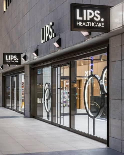
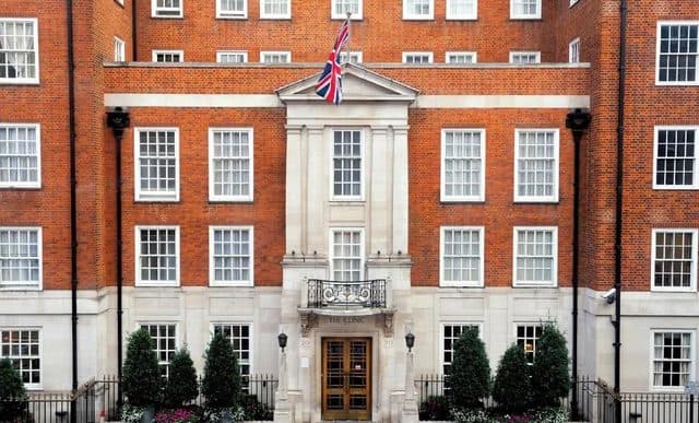
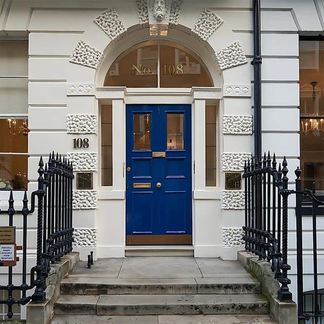
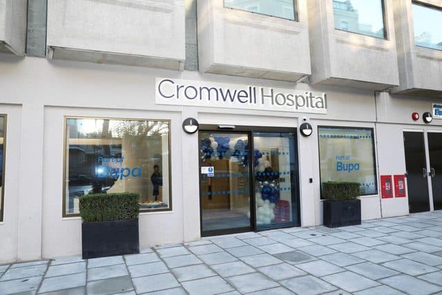
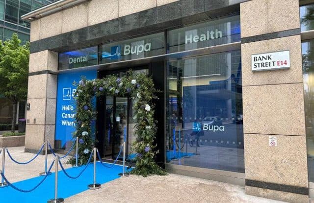
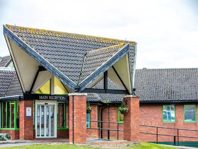
Read more: Request an appointment →Private consultations are available at 108 Harley Street, The London Clinic Rapid Diagnostics Centre on Harley Street, LIPS Healthcare at Battersea Power Station, BUPA at Canary Wharf and The Cromwell Hospital in London. The website’s Locations section contains current addresses and contact details. NHS appointments at Broomfield Hospital require an NHS referral.
Read more: Locations →Self-pay patients can usually arrange a private consultation without a GP referral. Insured patients should check their insurer’s rules, as many require pre-authorisation before the appointment or imaging and some require a GP referral. NHS appointments require the appropriate NHS referral pathway.
Dr Masannat is recognised by all major private medical insurers. Cover varies by policy, so obtain an authorisation number and confirm any excess, limits or restrictions directly with your insurer before attending. Bring the membership and authorisation details to the appointment.
Yes. Ask the team for the consultation fee and likely imaging or biopsy charges before the appointment. If surgery is recommended, you should receive a written estimate setting out the hospital, surgeon and anaesthetic fees and explaining which additional costs may apply.
Bring a list of medication and allergies, relevant clinic letters and pathology reports, and details of previous breast surgery or cancer treatment. If imaging was performed elsewhere, we will arrange for the original images—not only the written report—to be transferred in advance so please let the team know. Also bring your insurer’s authorisation details if applicable.
A video consultation may be useful for discussing an existing diagnosis, reviewing reports, obtaining a second opinion or planning next steps. New breast symptoms often still require an in-person examination and imaging, so a remote appointment may not replace a one-stop clinic visit.
Yes. International patients should send relevant reports and arrange secure transfer of imaging and pathology material before the appointment where possible. The team can advise whether an initial video review or an in-person one-stop assessment is more appropriate and can provide a proposed pathway and estimated costs.
No. The practice provides planned specialist breast assessment and treatment rather than an emergency service. For an urgent problem after an operation, use the contact instructions supplied by the treating hospital; for serious or life-threatening symptoms, call 999 or attend the nearest emergency department.
Question not here? Call +44 (0) 20 8194 8609 orsend a request →
Pick the clinic nearest you
Choose the clinic that suits you — 108 Harley Street is Dr Masannat's primary base.
108 Harley Street
Send a message to the team
For urgent concerns, please call — for everything else, this form reaches the front office directly.
Ready to talk to Dr Masannat’s team?
Reach the front office directly, or book a consultation.